Blog

New Cardiac Imaging Technique Produces Higher Quality Images in Less Time

CMR

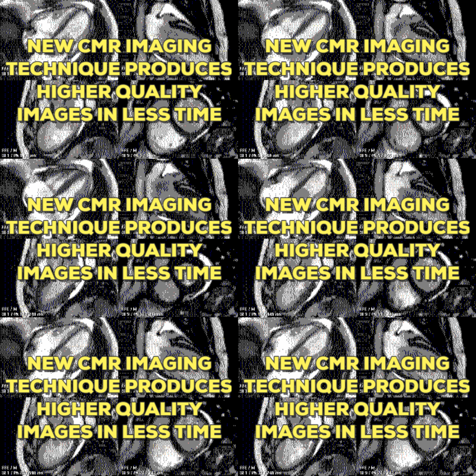
An innovation in cardiac magnetic resonance (CMR) imaging eliminates the need to correct images for respiratory motion, producing higher quality, more accurate images without waiting for patients to breathe.

Preliminary research presented at EuroCMR 2016 by Professor Juerg Schwitter, director of the Cardiac MR Centre at the University Hospital Lausanne, Switzerland, demonstrated how using a modified ventilator and small volumes of air, called "percussions," eliminated the need for patients to breathe during CMR.

Continue reading New Cardiac Imaging Technique Produces Higher Quality Images in Less Time

California Regulations to Relax on Fluoroscopy

In February, the Alliance of Cardiovascular Professionals updated members regarding progress made to remove a regulatory burden preventing provision of team-based care in California cardiac cath labs during procedures involving fluoroscopy.

In October, 2015, the Radiologic Technology Certification Committee in California unanimously passed a motion allowing an individual under the direct and immediate supervision of the S&O to use an assistant in the real time movement of the patient under fluoroscopy... (A Step Forward for CVTs in California, ACVP Advocacy)

On April 13th, 2016, the Radiologic Technology Certification Committee (RTCC) of California met with an agenda item to clarify this motion. The amended motion reads:

Continue reading California Regulations to Relax on Fluoroscopy

ICYMI: TAVR Out-Performs Surgical Replacement

This video from Diagnostic and Interventional Cardiology's Pulse TV shares the biggest piece of news from the American College of Cardiology's annual conference last month---that Transcatheter Aortic Valve Replacement (TAVR) out-performed surgical valve replacement in the PARTNER II trial.

Continue reading ICYMI: TAVR Out-Performs Surgical Replacement

VIDEO: Ultrasound identifies dangerous plaque

New research in ultrasound out of Lund University in Sweden might be key to better, broader screening for cardiovascular risk.

A relatively simple mathematical calculation developed at Lund University can be used to interpret ultrasound signals and identify whether or not plaques consist of harmless connective tissue and smooth muscle cells or dangerous lipids and macrophages.

Continue reading VIDEO: Ultrasound identifies dangerous plaque